test1
Flow & Mass Cytometry Core at the Stem Cell Research Center
Core Leadership and Personnel
The Flow Cytometry Core is led by Vanessa Scarfone, MSc, who has managed the facility since its inception in 2010 and brings nearly 20 years of experience in flow cytometry and cell sorting. Vanessa has extensive expertise in CyTOF experimental design, instrumentation operations, and high-dimensional data analysis, and has trained hundreds of researchers in advanced flow and mass cytometry techniques.
Faculty Advisor Dr. Matt Inlay brings over 25 years of experience using flow cytometry to study hematopoietic stem cells, embryonic development, and immune system interactions. His research employs transplantation and lineage tracing approaches, and he has served as the Core's faculty advisor for more than a decade.
Pauline Nguyen, Flow Cytometry Core Technician, has over 5 years of experience in flow cytometry and cell sorting. She supports daily operations, training, and technical assistance, ensuring optimal use of the facility's technologies and smooth coordination of experiments.
Contacts
- Director: Vanessa Scarfone, MSc – vanessa.s@uci.edu
- Faculty Advisor: Matt Inlay, PhD – minlay@uci.edu
- Core Technician: Pauline Nguyen – nguyenpu@hs.uci.edu
About the Core
The Flow Cytometry Core is a high-impact research resource, providing advanced single-cell and spatial analysis technologies essential for the development and characterization of human stem cell-based in vitro models. Our platforms enable deep phenotyping, rare cell detection, and precise cell isolation, supporting projects across basic, translational, and regenerative medicine research.
Our Core has contributed to over 100 peer-reviewed publications and supported more than $20 million in successful grant applications by providing preliminary data, experimental expertise, and letters of support. We train over 100 students, postdocs, and faculty annually, including the CIRM COMPASS Scholars, providing hands-on experience in the latest flow and mass cytometry technologies. In addition, we host regular seminars and technology workshops to ensure our research community remains at the forefront of methodological advances.
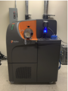
Our CyTOF mass cytometry system enables measurement of more than 50 parameters per cell, revealing detailed phenotypic and functional profiles of complex cell populations. BD flow cytometers and sorters facilitate population purification, single-cell cloning, and rigorous quality control to ensure reproducibility. The Hyperion Imaging Mass Cytometry platform adds spatial context, enabling the simultaneous detection of 40+ markers in tissue sections and organoids while preserving architecture and eliminating autofluorescence.
By integrating advanced instrumentation with expert guidance and a proven record of research success, the Flow Cytometry Core empowers investigators to produce high-quality, reproducible data that advances our understanding of human biology, disease mechanisms, and potential therapeutic targets.
Technologies
Mass Cytometry Systems
Flow Cytometry & Cell Sorting
Specialized Services
Workshops, Tech Talks, and Seminars
This course will cover the fundamentals of imaging mass cytometry, including its comparison with fluorescence-based spatial imaging, data dimensionality, experimental design, panel building, and sample preparation techniques like tissue processing and fixation. Participants will also learn about metal antibody conjugation protocols, titration, optimization, and data analysis using neural networks, segmentation, and dimensionality reduction. Students will work on antibody conjugation, sample preparation, and running samples.
This course focuses on foundational concepts, with lectures covering technical aspects of mass cytometry, its comparison to fluorescence-based cytometry, experimental design, data dimensionality, and panel building. Participants will learn about sample preparation techniques for surface and intracellular targets, as well as metal antibody conjugation protocols, titration, validation, and optimization. Students will engage in practical experience, followed by basic data analysis using tools like Cell Profiler, HistoCat, and advanced free software packages.