Seed Grant Awardees
Since the inception of the Seed Grant Program in 2014, we have awarded $454,000 in initial funding from private donors, family foundation and corporations. As a direct result of this initial funding, our researchers have been awarded an additional $11,285,000 in funding from larger agencies, including NIH, CIRM and others. That is a remarkable 25-fold return of investment. Our past Seed Grant awardees since the inception of the program are listed below.
Stem Cell-Derived Exosomes to Mitigate Alzheimer’s Disease Neuropathology
 Munjal Acharya, Ph.D.
Alzheimer’s disease (AD) is the most prevalent age-related neurodegenerative disorder, currently affecting more than 35 million people worldwide and estimates suggest that number will more than double by 2030. AD is typically characterized by the accumulation of A-beta plaques (ABPs) and neurofibrillary tangles (NFTs), which are accompanied by gliosis and widespread neuronal and synaptic loss, causing progressive loss of memory and cognitive function, and a steadily declining quality of life.
The Definition of Corneal Epithelial Stem Cells at a Single Cell Level
 Bogi Andersen, M.D.
 James Jester, Ph.D.
 Qing Nie, Ph.D.
We propose that newer methodologies—scRNA-seq and scATAC-seq--can be used to gain new insights into the differentiation states and heterogeneity of the CE. Specifically, we want to use these methods to identify CE stem cells with more precision than has been previously possible. We will use the genetically tractable mouse system to refine our approach, and then use that knowledge in the future studies into CE stem cells in human corneas. Our work is basic, but also translationally significant because it will ultimately give new insights into Limbal Stem Cell Deficiency, a common abnormality in corneal diseases.
A CRISPR-Cas9 Based Approach to Track Mitochondrial DNA Transfer and Investigate the Function of Donor hNSC in Cell-based Therapies
 Aileen Anderson, Ph.D.
Mitochondria are the center of mammalian energy production but also regulate other key cellular functions such as cell death and oxidative stress control. Mitochondria dysfunction is a hallmark of neurological disease and injury, including neurodegenerative disorders and aging1. Additionally, genetic mitochondrial disorders affect 1 in 5000 births in the USA.
In Situ Incorporation of Microglia During 3D Brain Organoid Development to Enhance Brain Development/Maturation
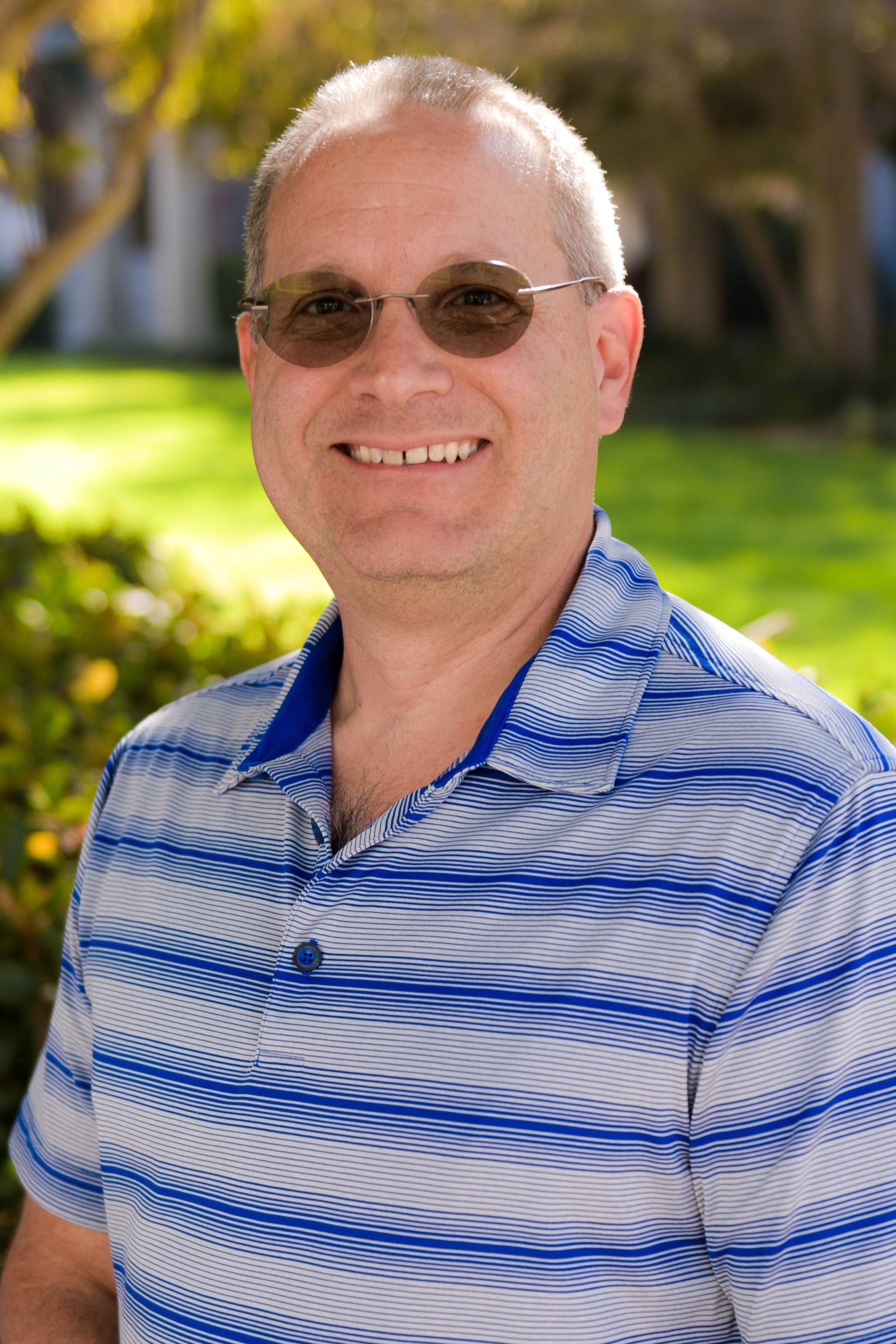 Brian Cummings, Ph.D.
 Anita Lakatos, Ph.D.
Traditional brain organoids lack microglia, a key cellular component of real brains. We seek to establish a human cortical organoid system that more realistically replicates human-specific brain development/maturation for brain research and drug discovery. While animal models can provide knowledge about neurodevelopment and neurodegenerative diseases, their translational value is limited due to the complexity of the human brain and unique proteins expressed in human tissue (e.g. ApoE, As, or tau). Progress in the development of 3D organoids from pluripotent stem cells has created an unprecedented opportunity to gain insight into human neurodevelopment and complex brain function to complement animal studies.
Dual Calcium Activity and Morphological Reporter Line for the Study of Human Cell Types
 Sunhil P. Gandhi, Ph.D.
When encountering injury-associated debris or pathogens, microglia in the nervous system mount a highly localized and finely calibrated inflammatory response. State-of-the-art in vitro models do not adequately capture the spectrum of human microglia responses found in vivo; new tools are needed to interrogate human microglial activity in response to specific insults in the living brain. We propose to develop a new experimental platform that uniquely combines recent developments from our research groups in human microglial stem cell technology and xenotransplantation into mice (Blurton-Jones), in vivo multiphoton imaging (Gandhi), and a recently created genetically encoded, dual indicator of calcium activity and cell morphology, Salsa6f (Cahalan). Specifically, we propose to create and validate a Salsa6f calcium/morphology indicator human iPS cell line using CRISPR technology. The Salsa reporter cell line would be the first of its kind for a human iPSC system. Once validated for microglia cell studies, we envision the Salsa6f cell technology will be used to investigate calcium activity in a vast array of iPSC-derived human cell types.
Suppression of Keloid Scars by Inactivation of MEST in Fibroblasts
 Maksim Plikus, Ph.D.
We will investigate new therapeutic approach to keloid scarring aimed at converting proliferative fibroblasts into non-dividing myofibroblasts by turning off the abnormally-activated keloidspecific signaling mechanism. This approach, if works, will effectively suppress uncontrollable growth of keloids without the need for surgery and radiation. We propose that MEST (Mesoderm Specific Transcript) is the abnormally overactivated signal in keloid fibroblasts, “locking” them in the state of proliferation and preventing differentiation toward myofibroblasts. Inactivating MEST or its activity will break the pathological signaling, enabling keloid fibroblasts to differentiate. MEST is a member of α-β hydrolase protein family.
Generation of TurboID-HTT isogenic iPSCs via CRISPR genome editing to study protein-protein interactions in Huntington’s disease
 Leslie Thompson, Ph.D.
Since its discovery, mutated Huntington (mHTT) has been implicated in multiple pathways including dysregulation of protein scaffolding and autophagy.1-6 The complete elucidation of HTT’s function will significantly enable therapeutic interventions. Understanding how HTT interacts with other proteins and RNAs is crucial to the further elucidation of its’ function(s) both normally and in disease. Recently, a major focus of the neurodegeneration field has been to understand the role of stress granules (SGs) in neurotoxicity.7,8 Interestingly, HTT interacts with core SG proteins, Caprin1 and G3BP1.9 Furthermore, HTT interacts with valosin-containing protein (VCP) and the chaperonin protein TRiC/CCT, important SG regulators.10,11 Here we hypothesize that HD patients have altered SG dynamics due to (m)HTT’s ability to interact with core SG proteins. Our preliminary findings show that there are less SGs in iPSC-derived neurons from HD patients compared to control subjects as a result of heat-shock.
Small Vessels, Big Problems: The Role of MGP and Alk1 in Vascular Malformations
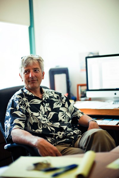 Christopher Hughes, Ph.D.
Matrix Gla-protein (MGP) is a vitamin K dependent protein locally produced in the vasculature. Both endothelial cells (ECs) and smooth muscle cells (SMCs) are able to synthesise MGP. To be functionally active, MGP needs to undergo post translational carboxylation which is driven by its cofactor vitamin K. MGP deficiency in SMCs results in extensive calcification of large and medium sized vessels, both in rodents and humans. The use of vitamin K-antagonist, thereby inducing a drug-induced MGP inactivity, also results in vascular calcification.
Visualizing the Emergence of Hematopoietic Stem Cells by Time-Lapse Imaging of Mouse Embryo Cultures
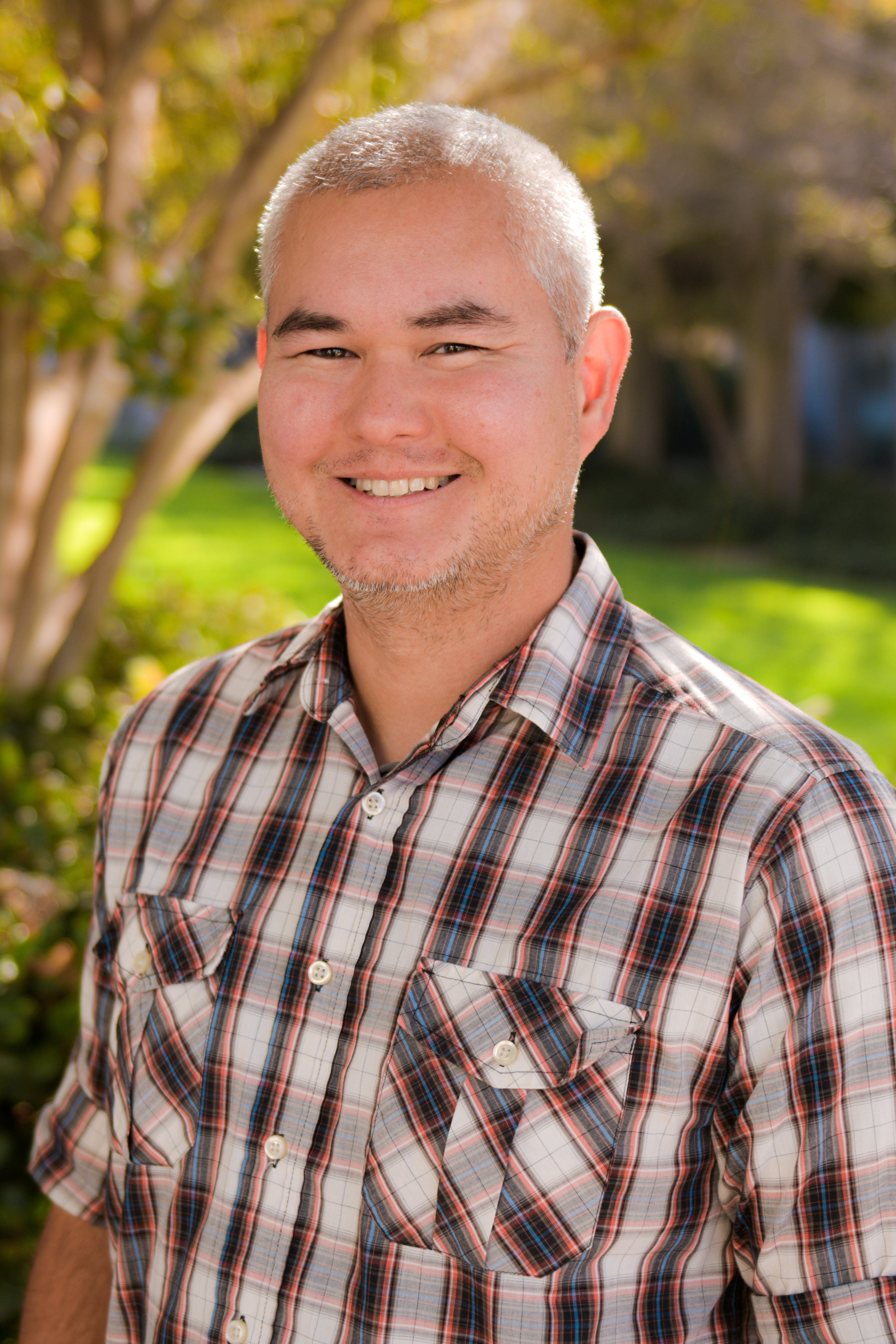 Matthew A. Inlay, Ph.D.
Laying dormant within our bone marrow is a type of stem cell that can produce our entire blood system, which includes all the protective cells of our immune system. These blood-forming stem cells are the key players in life saving bone marrow transplants. If taken from a healthy donor, a transplant of bone marrow can completely replace a patient’s diseased blood system, offering a potential cure for nearly any blood disease. Despite this potential, bone marrow transplantation is a last resort treatment. This is because of dangerous complications that can occur, such as opportunistic infection or graft failure. Pluripotent stem cell technology could be used to generate a limitless supply of blood-forming stem cells to reduce these complications, but this has yet to be achieved. In order to create new blood stem cells, we must first understand how blood stem cells are created naturally during the course of embryonic development. There is much we do not know about how the first blood stem cells arise in the embryo. We have evidence that a membrane that surrounds the embryo body, called the “yolk sac”, contains special blood vessel cells that directly produce the first blood stem cells. We have proposed to use mouse embryo cultures, and a new state-of-the-art microscope recently acquired by the Stem Cell Research Center, to directly observe the birth of blood stem cells in the yolk sac. If successful, our research will prove where blood stem cells come from, and allow us to focus on the yolk sac to better understand how this process occurs. By doing so, we could then replicate this in a dish to create new blood stem cells that could be safely used to treat blood diseases.
Drug Screening on Neurons with Epilepsy-Related SCN1A Mutations by Monitoring Large-Scale Network Activity with Multi-Electrode Arrays
 Diane O’Dowd, Ph.D.
Individuals with epilepsy, including GEFS+ and the more severe disorder Dravet Syndrome (DS), have seizures caused by uncontrolled electrical activity in the brain. Genetic analysis of many families demonstrates that both GEFS+ and DS can be caused by mutations that can occur at different locations in a specific gene encoding a sodium channel protein. This channel opens and closes, regulating the flow of sodium into brain cells that is critical for brain cell activity. The symptoms and responsiveness to drugs varies widely in individuals with both GEFS+ and DS. This is likely to be due to mutations at different locations in the gene causing distinct types of channel dysfunction, such as staying open too long or making it harder to open. To develop more effective drugs, it is important to know what can go wrong with the sodium channel in different patients and how this affects activity in single brain cells and cell networks. This is difficult to determine because monitoring activity of sodium channels in brain cells of people is not yet possible. The goal of this project is to use a new tool, known as multi-electrode arrays (MEAs), to measure the electrical activity of multiple human brain cells at the same time. Our lab has developed a rapid and reproducible way to turn human stem cells, originating from patient skin cells, into brain cells that can grow in a petri dish. This allows us to directly examine how sodium channels open and close, and how this regulates electrical activity in a human brain cell. We have generated stem cells from patients with specific epilepsy mutations. In addition, when we do not have access to patient cell lines, we have introduced specific mutations into control stem cell lines which are generated from individuals that don’t have epilepsy. By monitoring individual cells with classic recording techniques, we have shown that some mutations disrupt brain cell activity by altering the ability of the channel to open and others that prevent normal channel closing. Since seizure episodes in patients are caused by uncontrolled activity of groups of brain cells, the ability to evaluate activity in multiple cells at once with the MEAs will provide important new insights into the mechanisms of seizure generation. In addition, it will be much easier to screen for compounds that counteract abnormal network activity associated with patient specific mutations than our current method of recording activity in single cells.
Development of the Cancer Stem Cell-Driven Breast Cancer Animal Models for Single-cell Resolution Lineage Tracing
 Olga V. Razorenova, Ph.D.
Obtaining cell ancestry (lineage) in a multicellular organism is critical to our understanding of normal organ development, as well as diseases of development and cancer. Since the discovery in 2003 of cancer stem cells (CSCs) being the main contributors to tumor growth, there has been a long-standing interest in determining the lineage of individual CSCs to understand their roles in promoting tumor growth and metastasis. We are collaborating with Dr. Chang Liu here at UCI, who is developing a novel lineage tracing technology based on mutation-based rapid evolution of an artificial DNA fragment, which will allow to accurately determine the lineage of a single CSC in the primary tumor and metastases. Thus, the long-term goal of this research project is to answer three basic cancer biology questions and more: 1) Do metastases arise from closely or distantly related cells in the primary tumor? 2) Is there a constant exchange of cells between metastases and the primary tumor? 3) How many cell divisions should a CSC undergo to become a metastasis initiating cell? Although these questions are likely general to most solid tumors, the research will be conducted in triple-negative breast cancer (TNBC) mouse models. This pilot grant will allow us to select mouse models of TNBC, which rely on CSCs for tumor growth and metastasis and metastasize with high frequency to the lungs. These models will be suitable for experiments to determine the lineage of single CSCs, which will aid answering the above questions when a larger funding source is secured..
Cardiac Lineage Commitment Through Epigenome Manipulation
 Tim Downing, Ph.D.
Heart failure remains a leading cause of death in both adults and children, affecting over 14 million people worldwide. Given that the heart does not have the ability to regenerate on its own, we turn to regenerative medicine to restore lost cardiac function. Our research is aimed at developing new technologies to help differentiate human stem cells into beating heart cells (e.g., cardiomyocytes). After a heart attack these cells die or lose their ability to function properly making it difficult for the heart to pump sufficient amounts of blood. Furthermore, there is a pressing need for patient specific cardiomyocytes to be available for drug screening. However, current strategies to produce cardiomyocytes from stem cells yield cells that are not mature enough for therapy or drug screening. We hope to address this unmet need through the development of a technology that precisely controls epigenetic modifications within stem cells and cardiac fibroblasts in order to facilitate their transformation into cardiomyocytes that will be more suitable for use in regenerative medicine.
Characterizing the Impact of a DRAK2 Deficiency on Regulatory T-Cell Induction by Neural Precursor Cells
 Craig Walsh, Ph.D.
 Peter J. Donovan, Ph.D.
The overarching goal of this application is to characterize the influence of regulatory T cells (Tregs) on the outcome of stem cell based therapies to promote remyelination in diseases such as multiple sclerosis (MS). We have demonstrated that intraspinal transplantation of neural precursor cells (NPCs) derived from human embryonic stem cell (hESC) and induced pluripotent stem cell (hiPSC) lines leads to marked remyelination and neurologic repair following intracranial infection of mice with murine hepatitis (MHV).
Modeling Pediatric TBI with Stem Cell-Derived Brain Organoids & Laser Cavitation Bubble
 Brian Cummings, Ph.D.
Our goal is to use borganoids to develop a controlled in vitro model of TBI, in which panels of biomarkers, therapeutic candidates, and genetic risk factors can be screened with higher throughput than can be achieved in animal models. Using human ES cells, we can grow multiple, genetically identical organoids, subject them to precisely controlled injuries, return them to culture and subsequently track the development of neuropathology, screen for biomarkers of injury, and/or treat the injured borganoids with therapeutic compounds to assess injury prevention.
A Stem Cell Derived Therapy for Mucopolysaccharidosis Type I in Children
 Edwin S. Monuki, M.D., Ph.D.
Embryonic stem cell-derived CPECs (dCPEC) offer the possibility of a treatment that would otherwise be unattainable. CPECs have little replication in culture [2], and cannot be obtained in substantial quantities from donors. Our method for deriving dCPECs from embryonic stem cells (ESC) enables large-scale production [3, 4] and makes preclinical testing and disease treatments feasible. When transplanted into the ventricles, CPEC or dCPEC engraft into the host choroid plexus (Fig. 1 and [3]). These cells are highly secretory, long-lived, and exhibit no appreciable turnover. This greatly reduces risks of neural circuit disruption/seizures, unwanted differentiation, and tumorigenicity. Therefore, dCPECs are ideal for natural, safe, and long-term secretion of therapeutic proteins into the CNS.
Identifying a Role for Hedgehog Signaling in Hematopoietic Stem Cell Emergence in Embryonic Development
 Matthew A. Inlay, Ph.D.
The proposed study seeks to establish a novel regulator of HSC emergence in embryonic development. The data generated could provide powerful preliminary data that could be used for a larger grant, as no one has conclusively connected Hh signaling to HSC emergence. Furthermore, time-lapse imaging of HSC emergence in a living intact mouse embryo has yet to be reported. By identifying a role for Hh signaling in driving the formation of the first pre-HSCs, we hope to utilize this pathway to generate HSCs from pluripotent stem cells. This will help overcome that developmental roadblock. If successful, this could allow us to produce an unlimited number of pure, patient-specific HSCs, which could make BMT a completely safe and effective therapy without risk of graft failure, rejection, or GVHD for patients.